FETAL PIG DISSECTION MANUAL
A series of anatomical illustrations created for Arcadia University’s Biology Department.
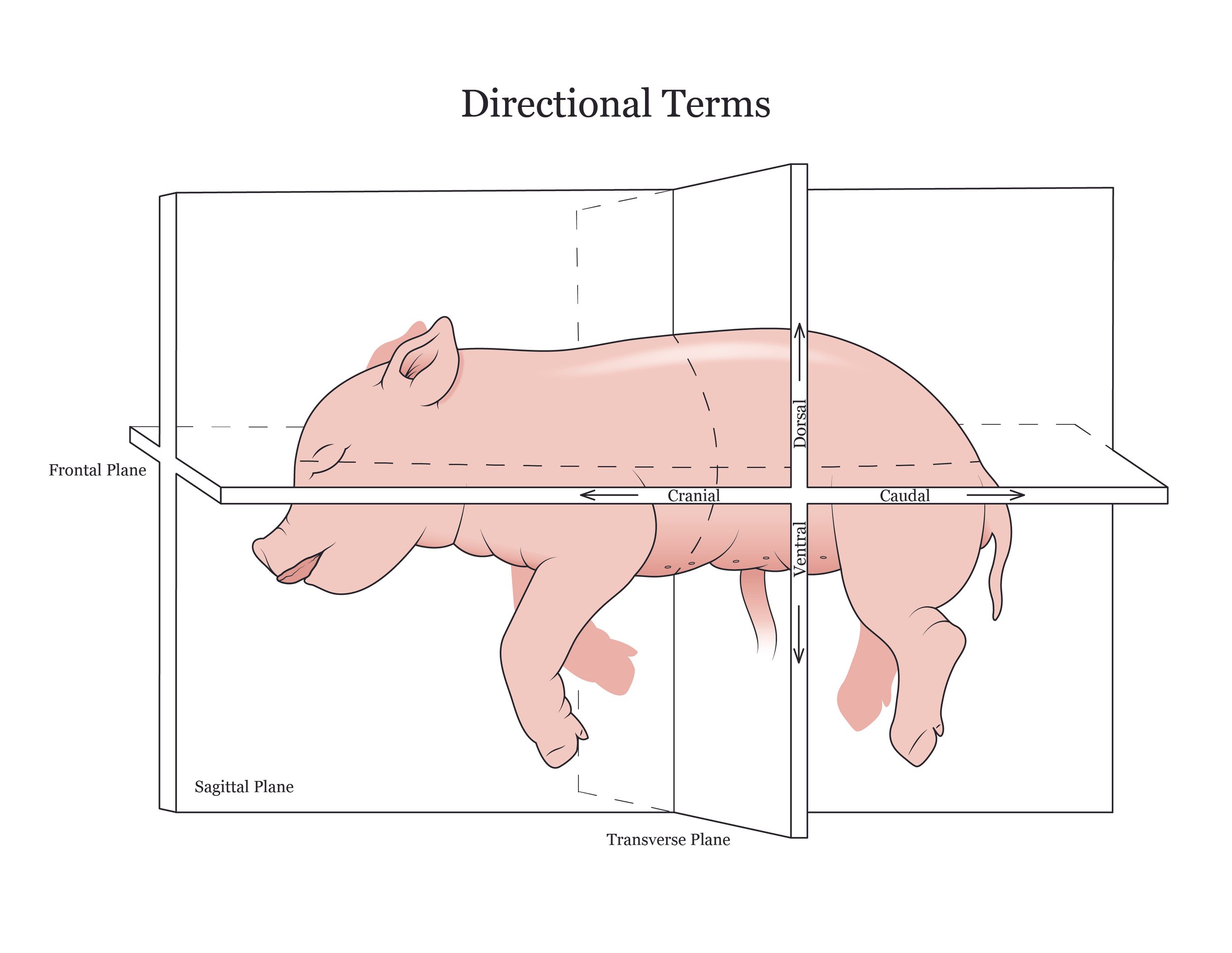
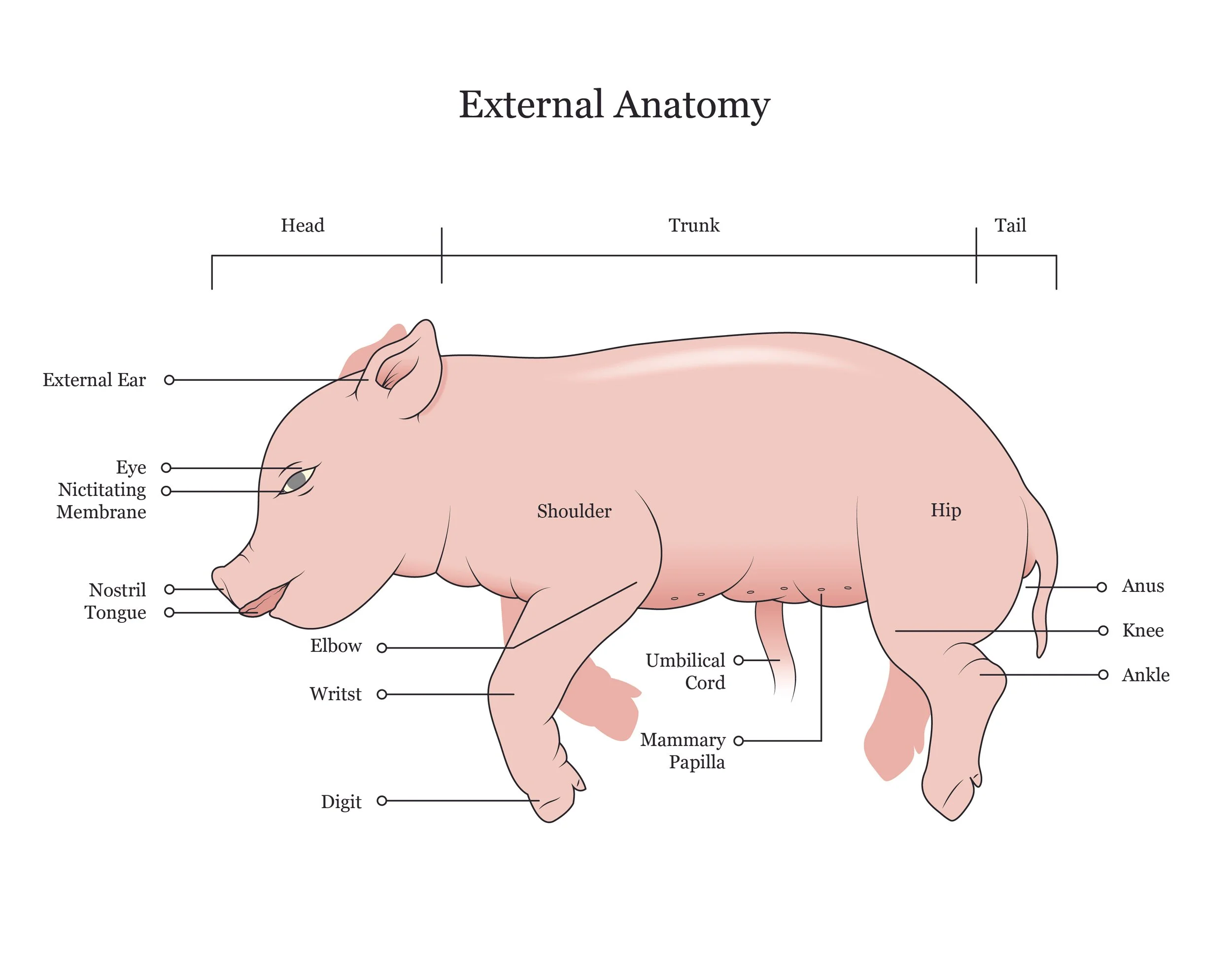
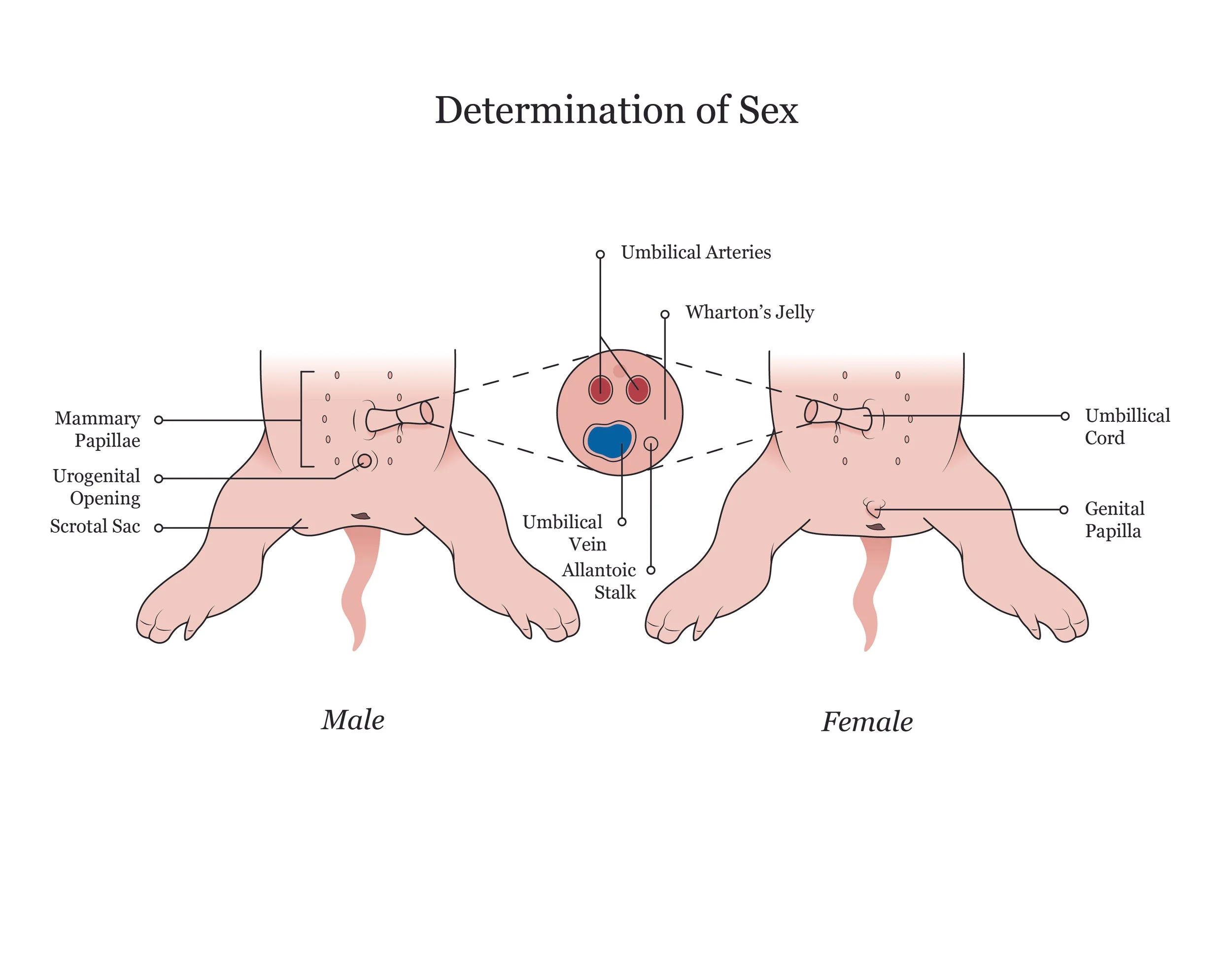
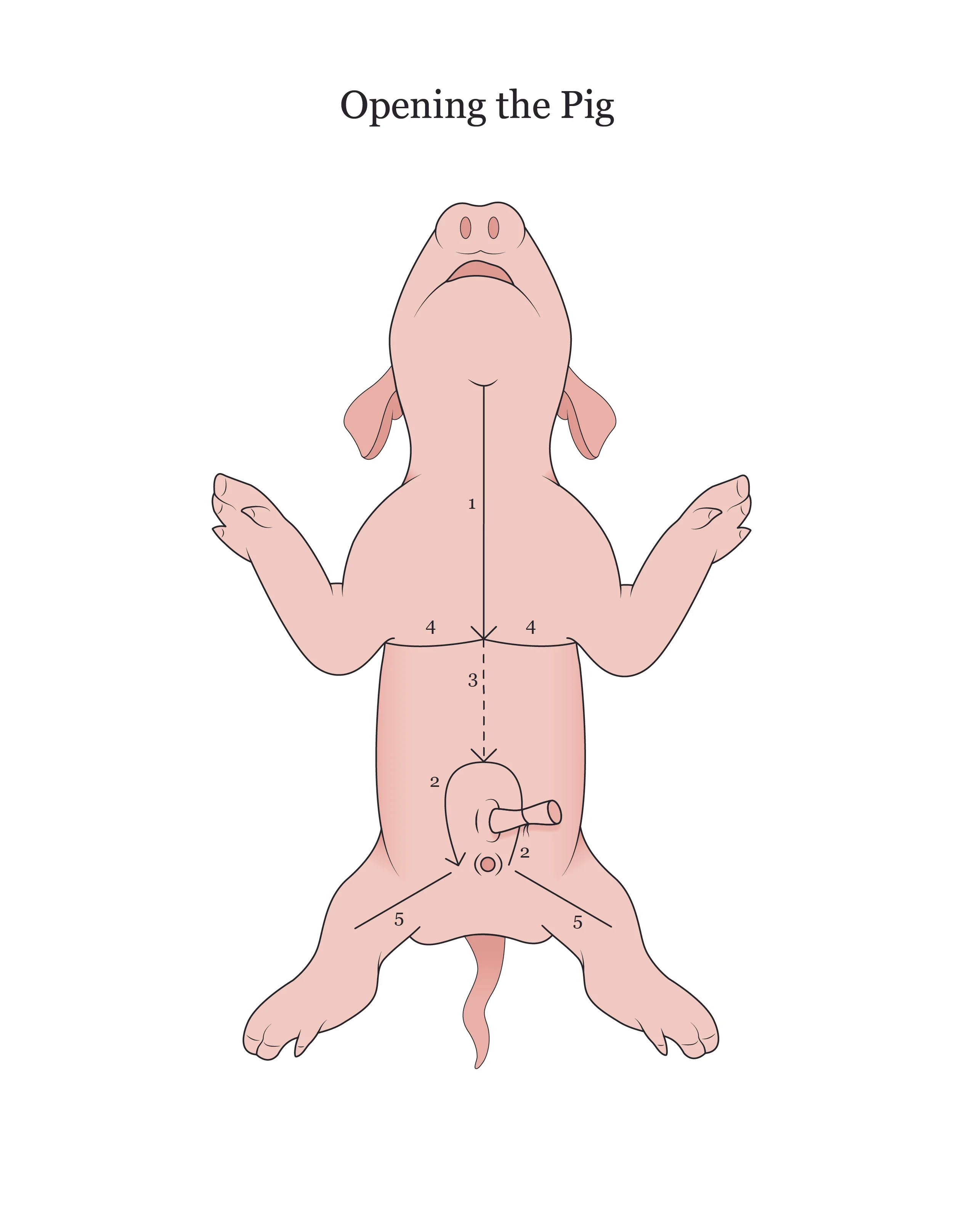
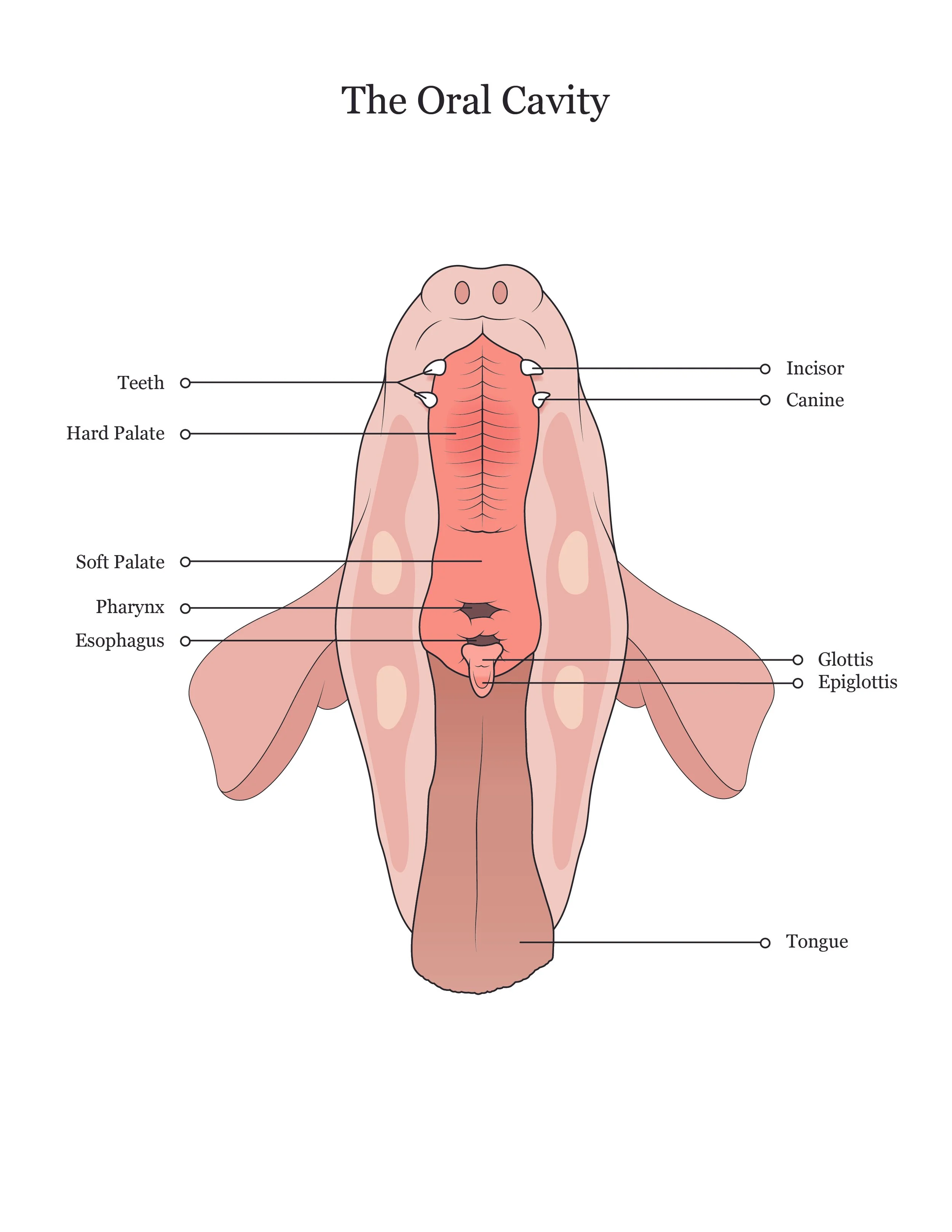
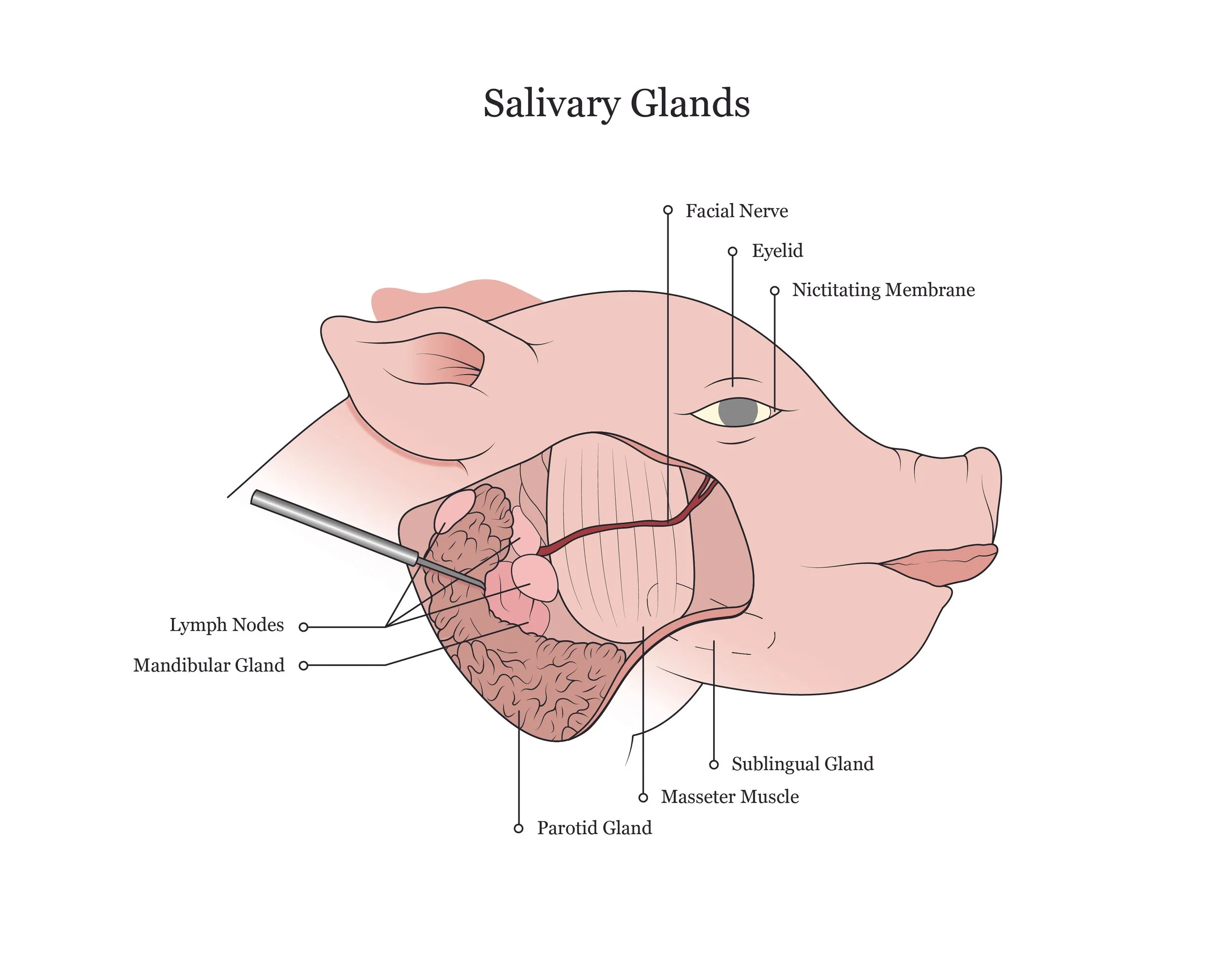
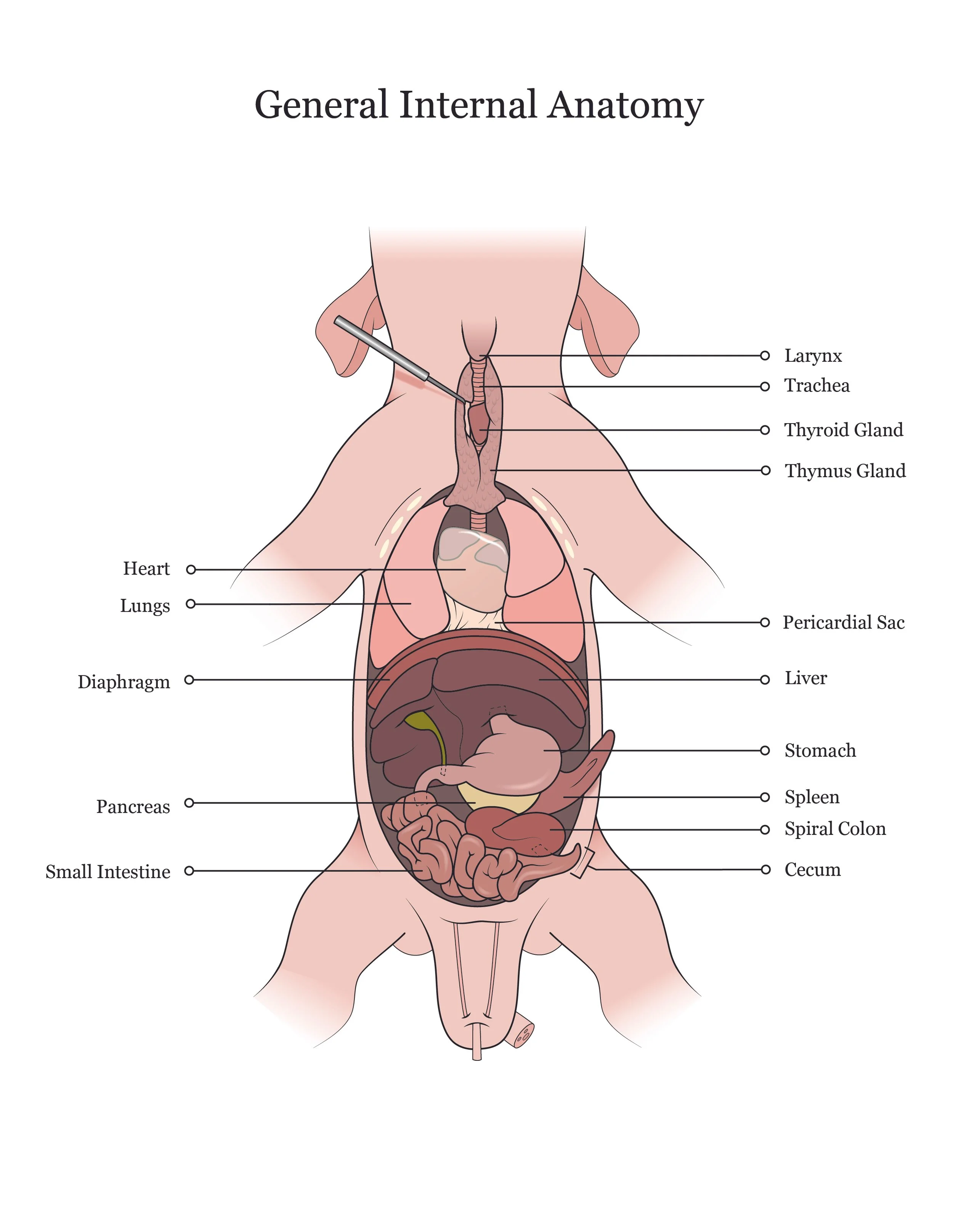
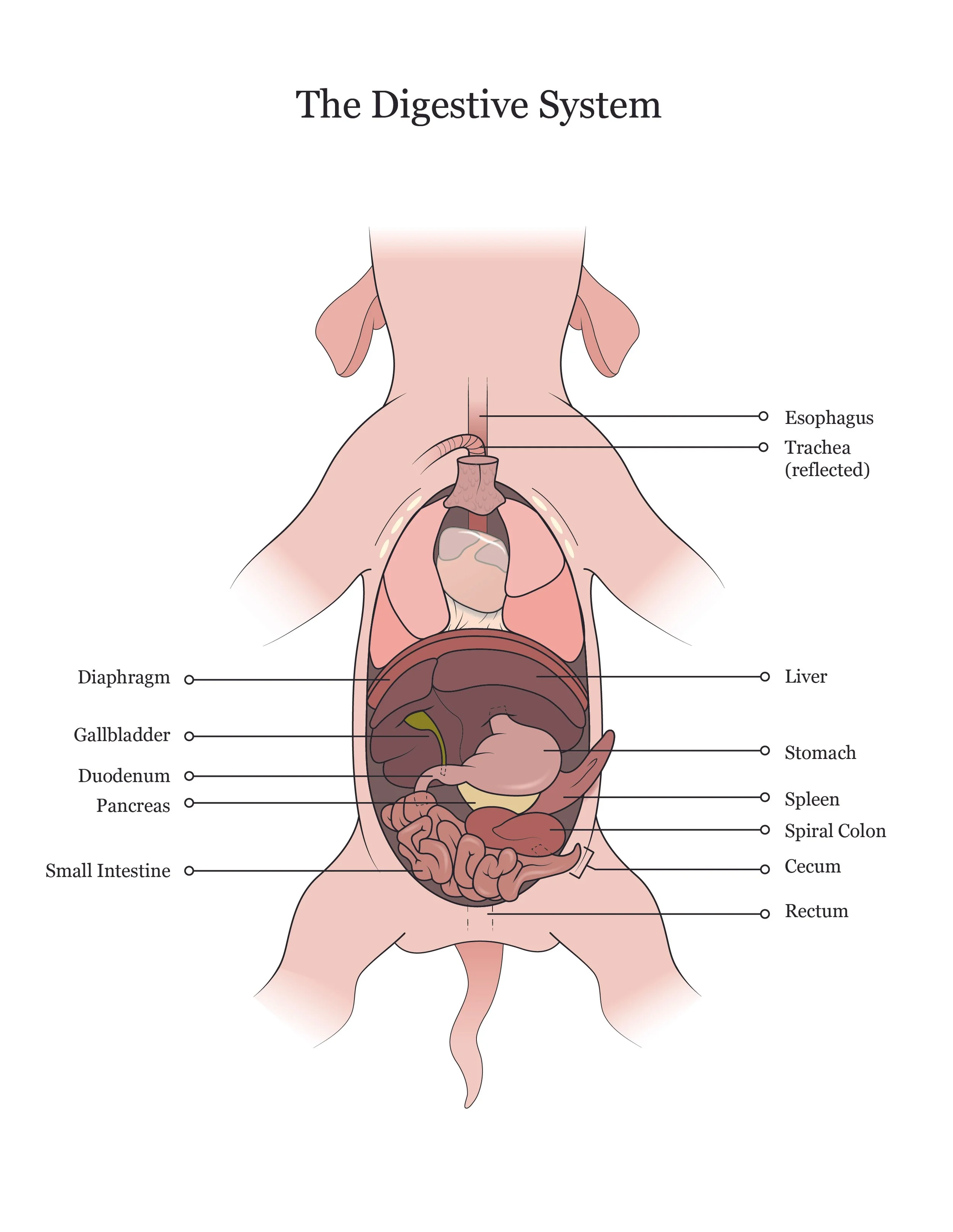
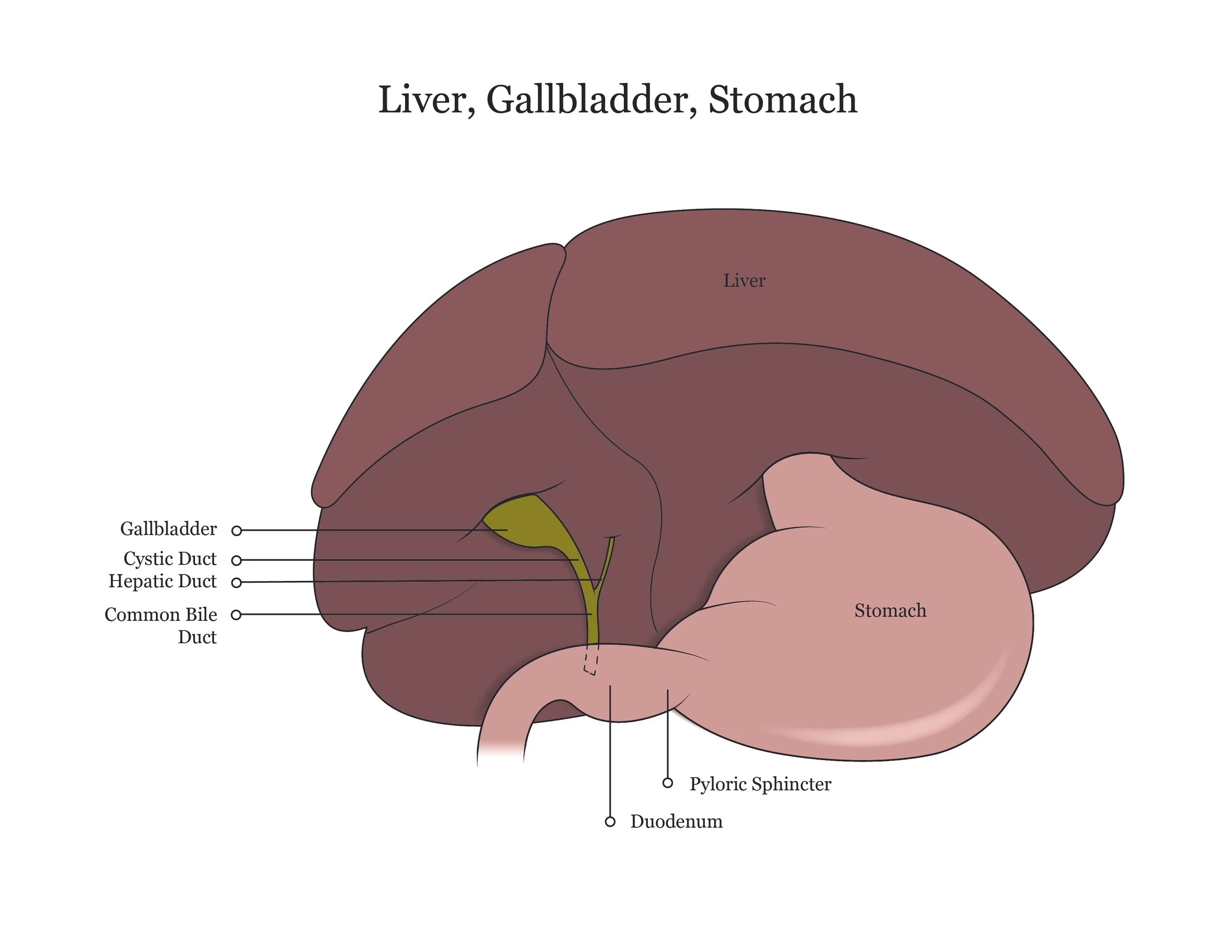
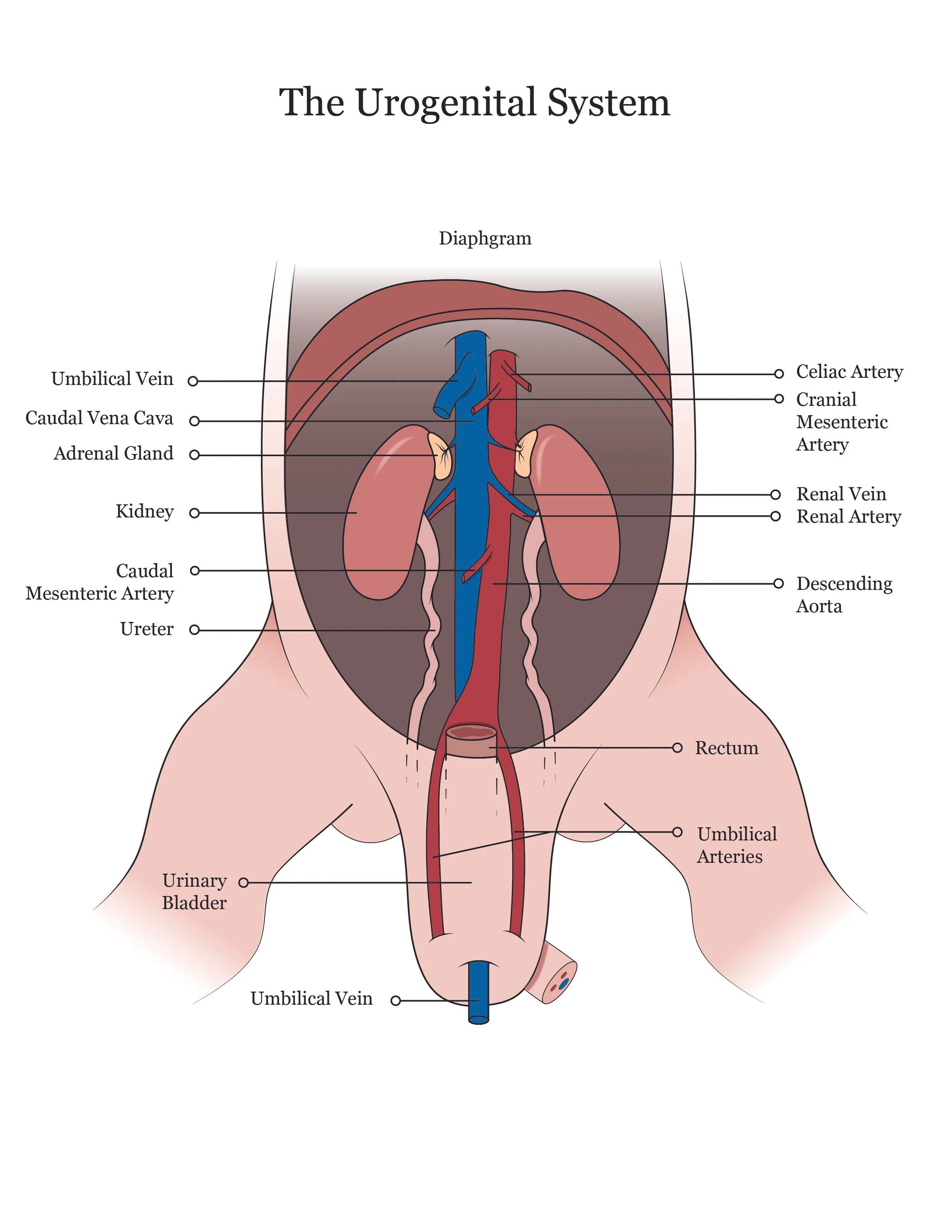
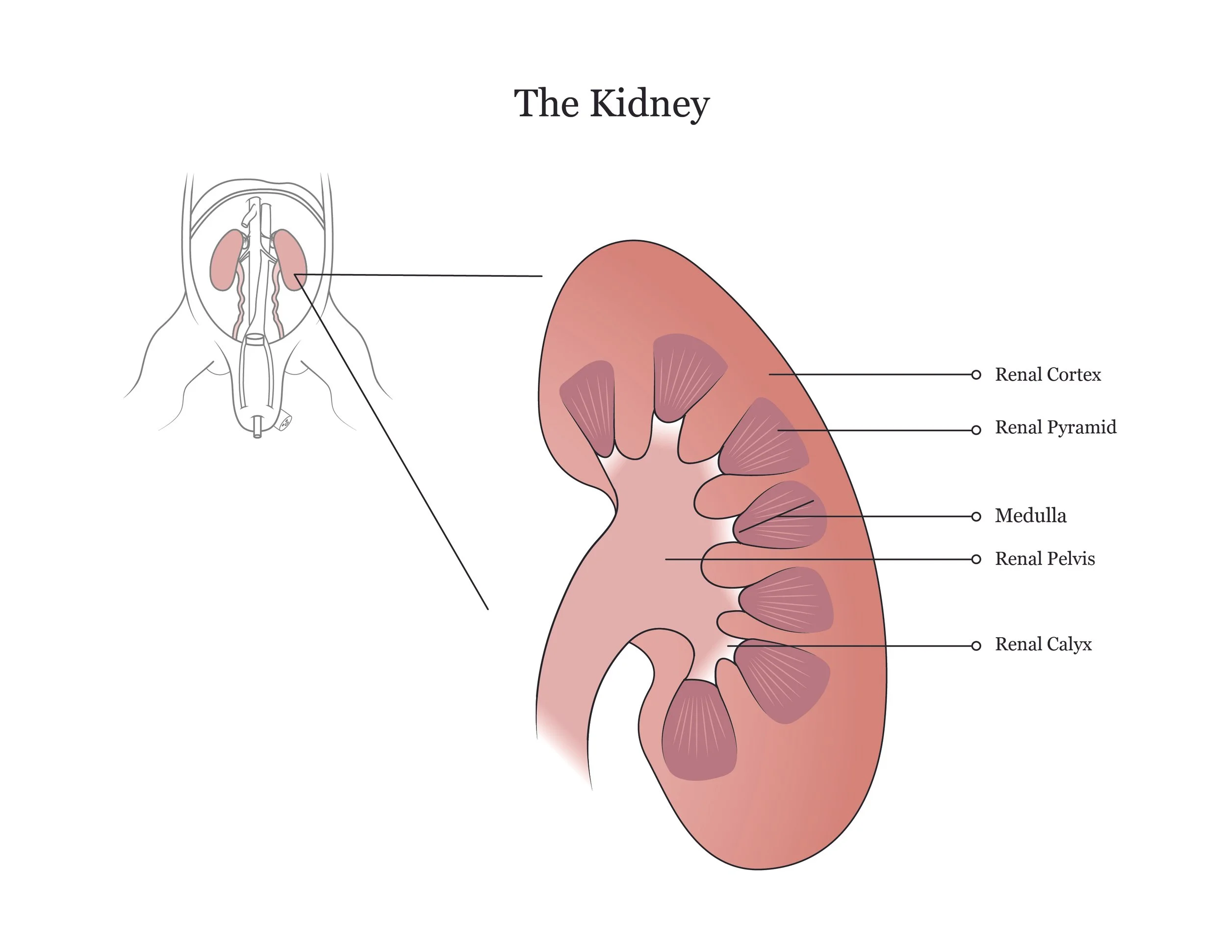
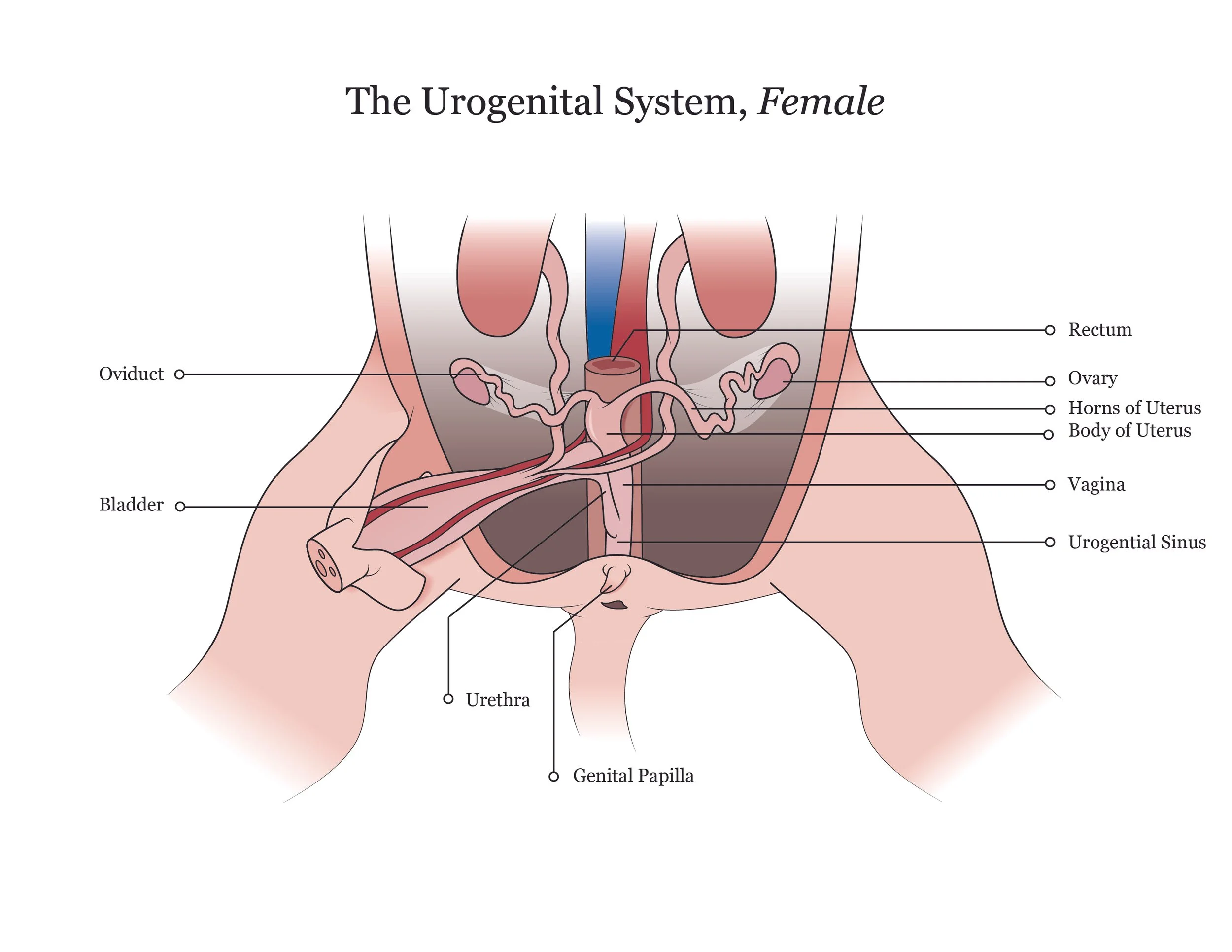
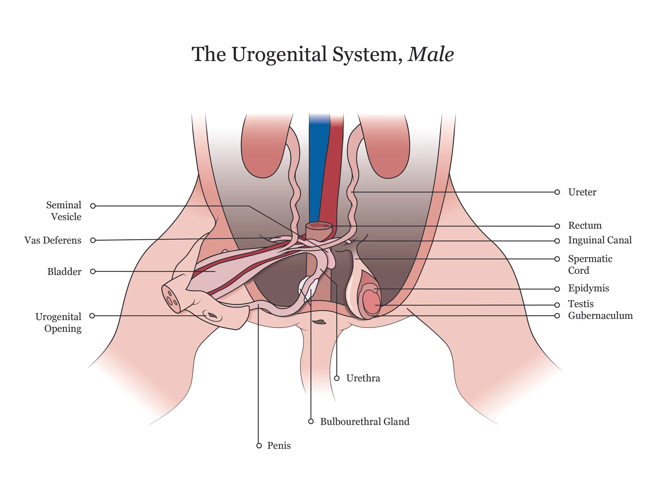
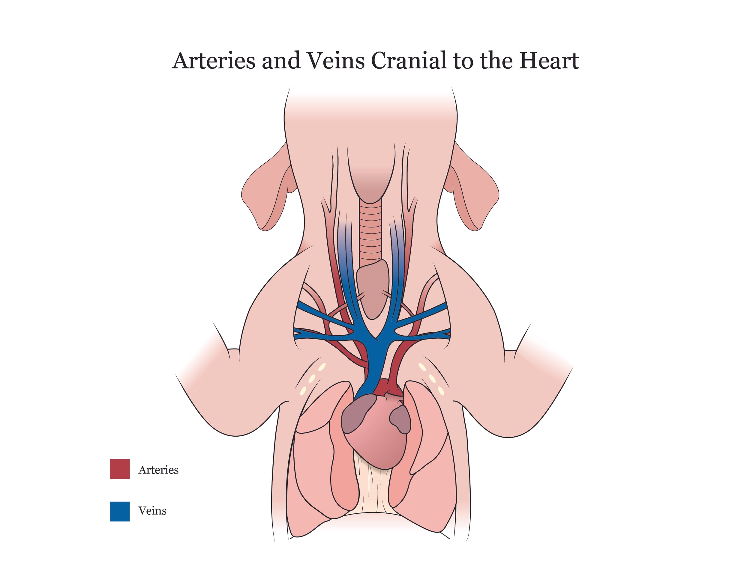
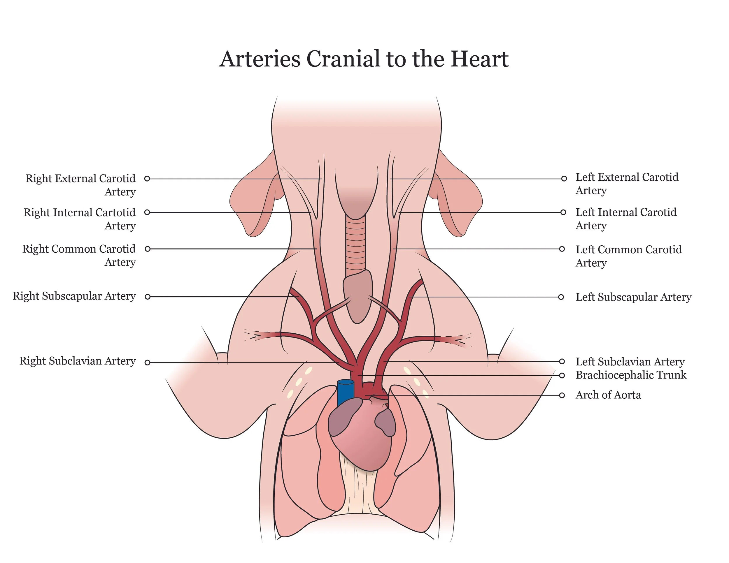
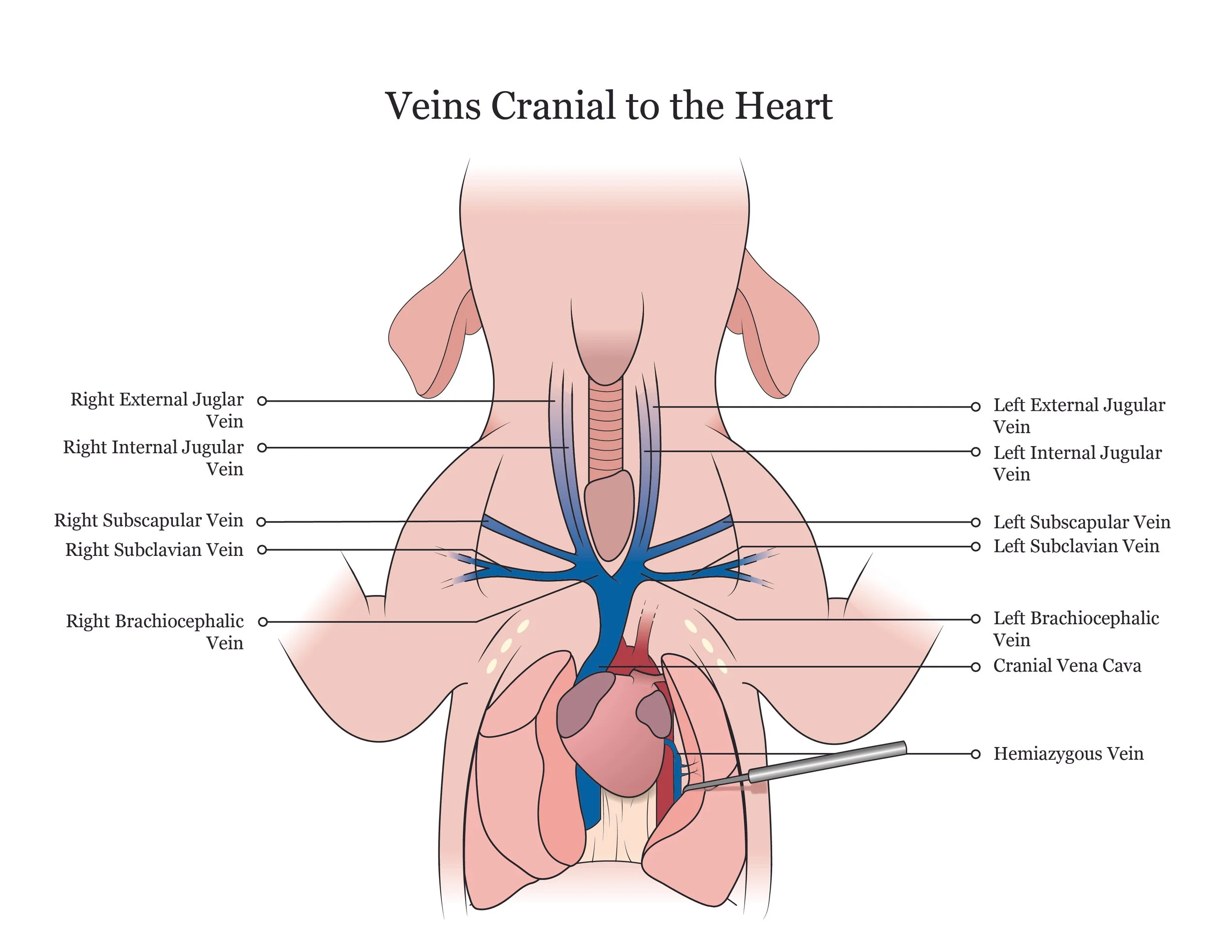
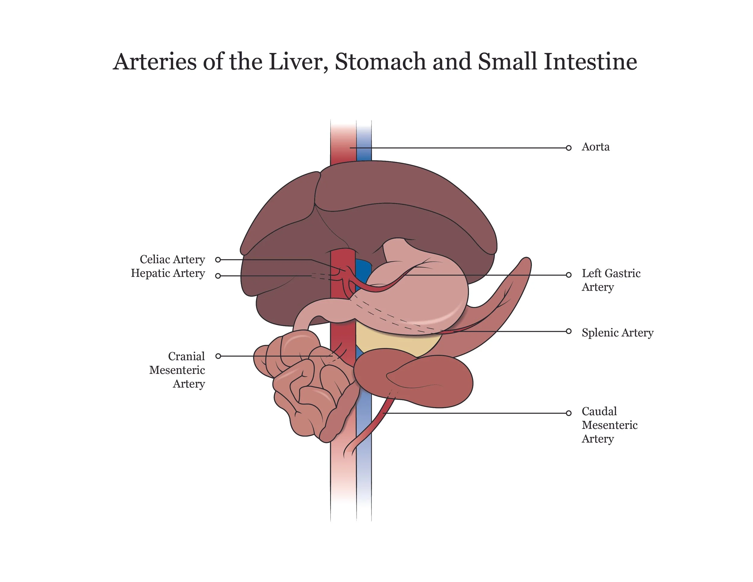
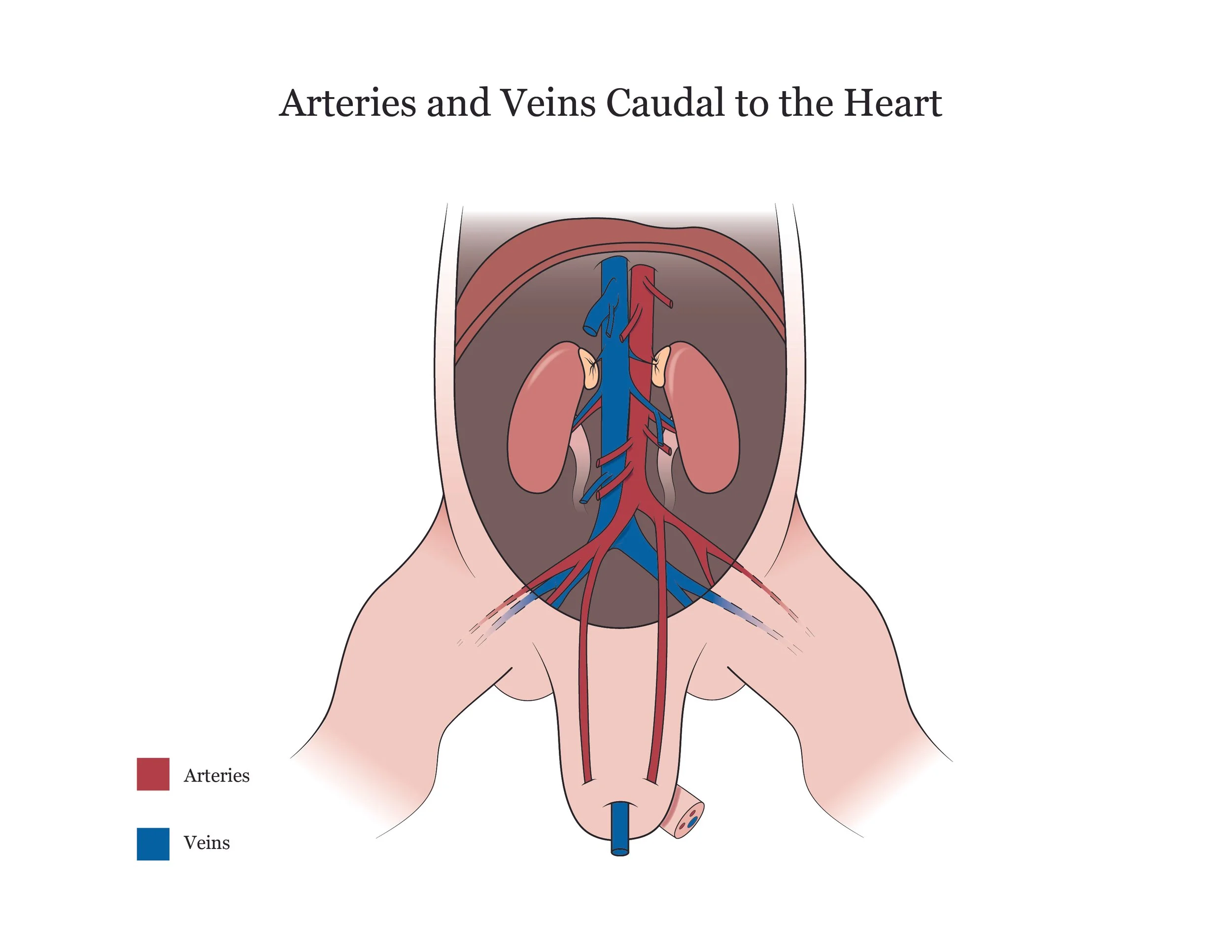
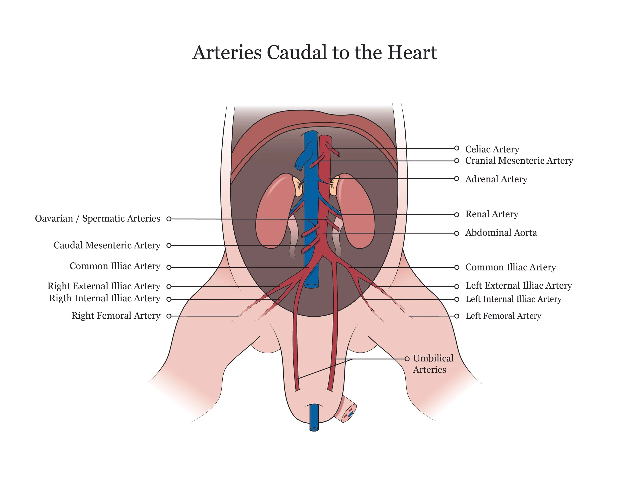
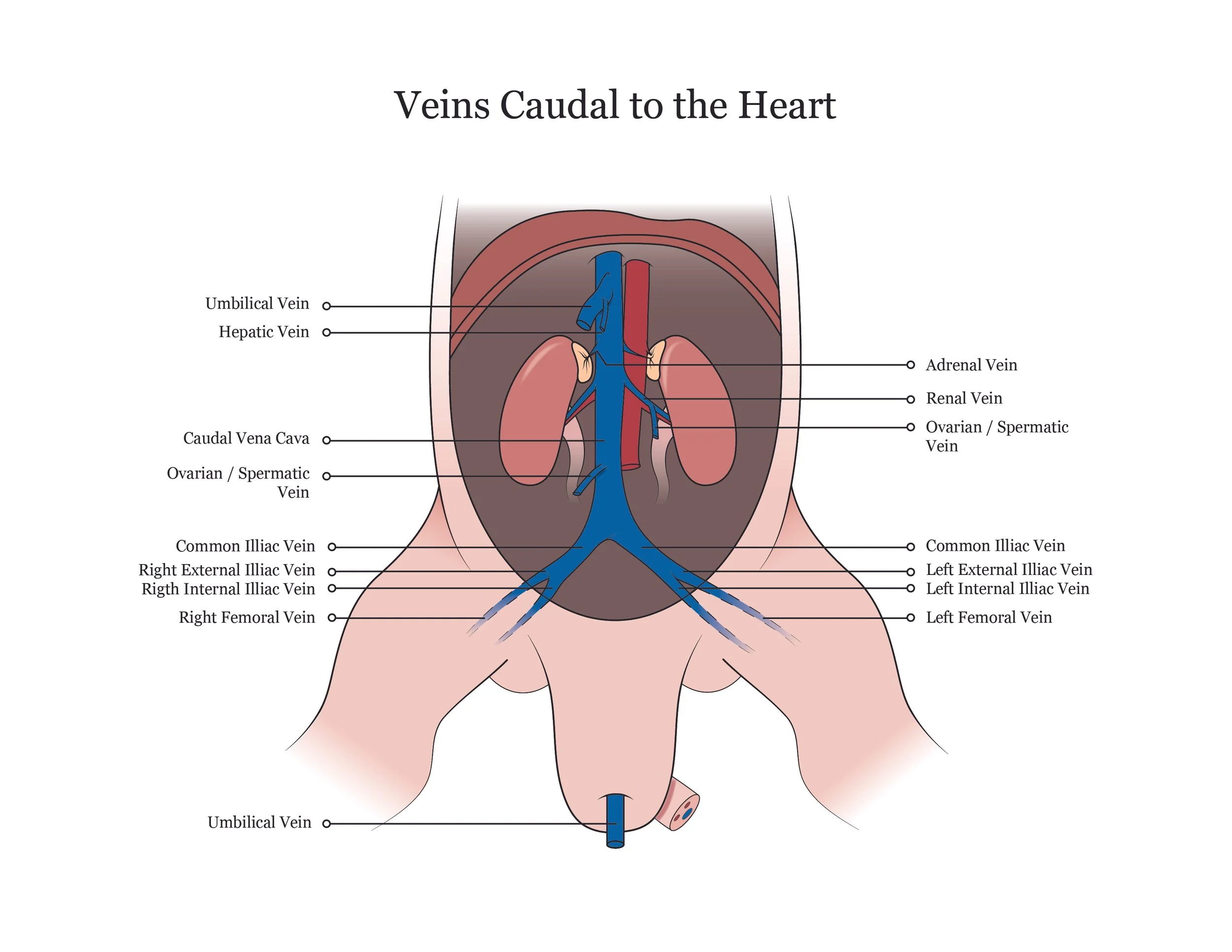
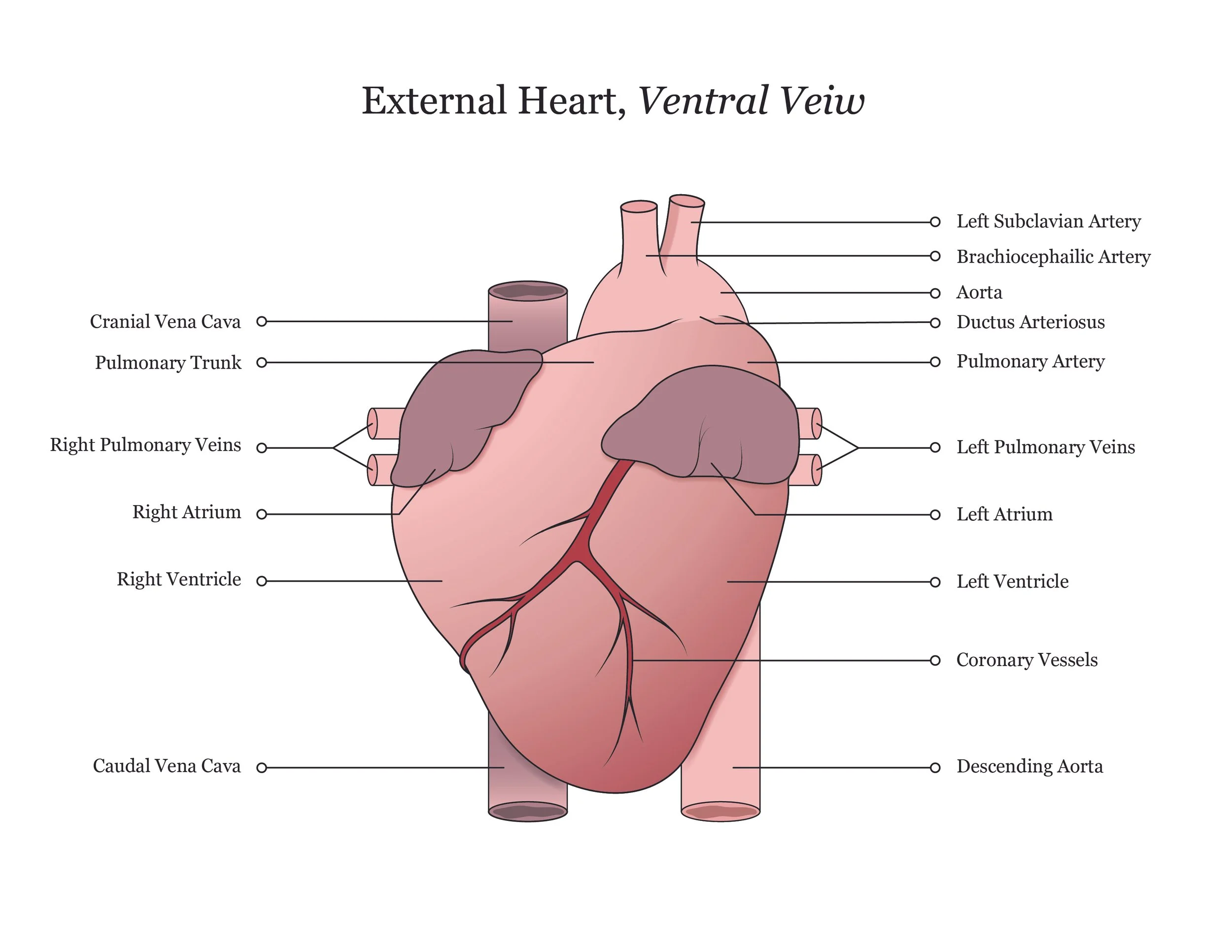
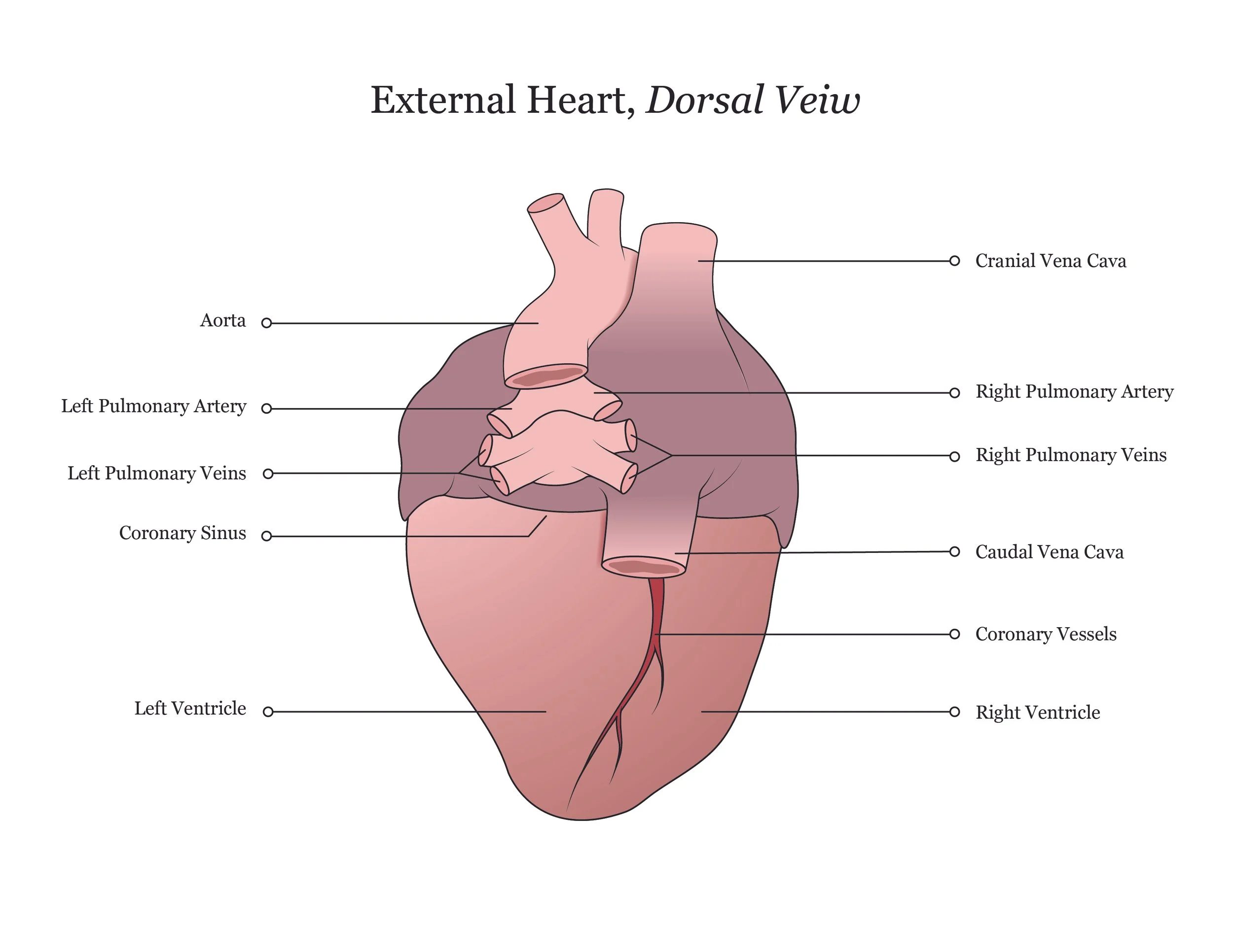
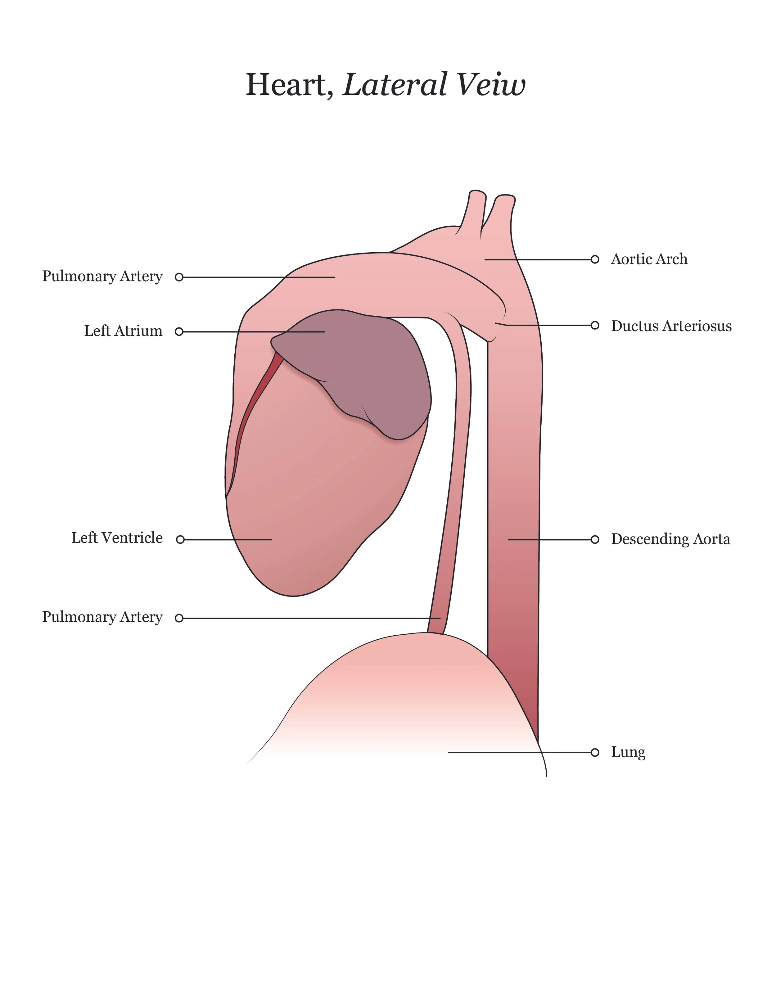
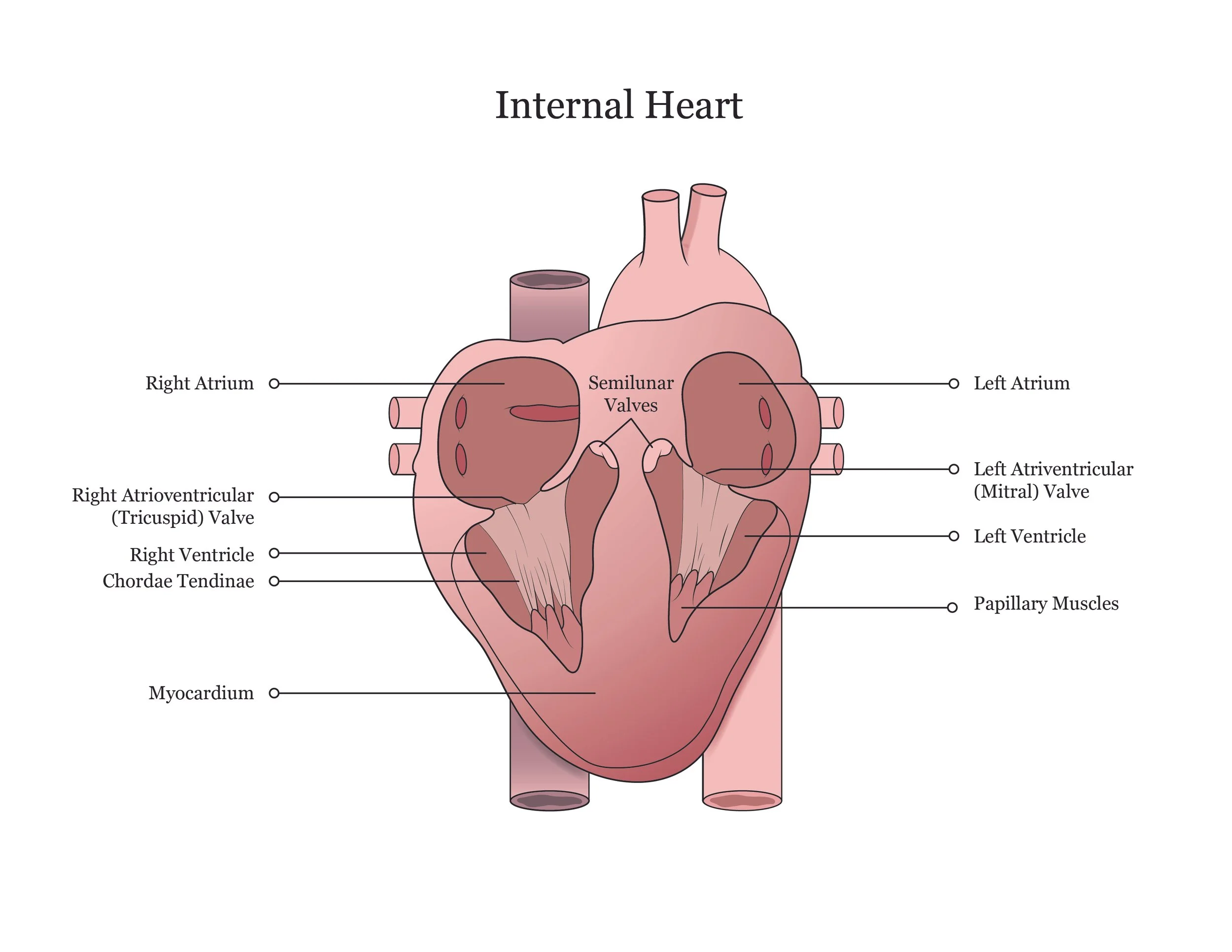
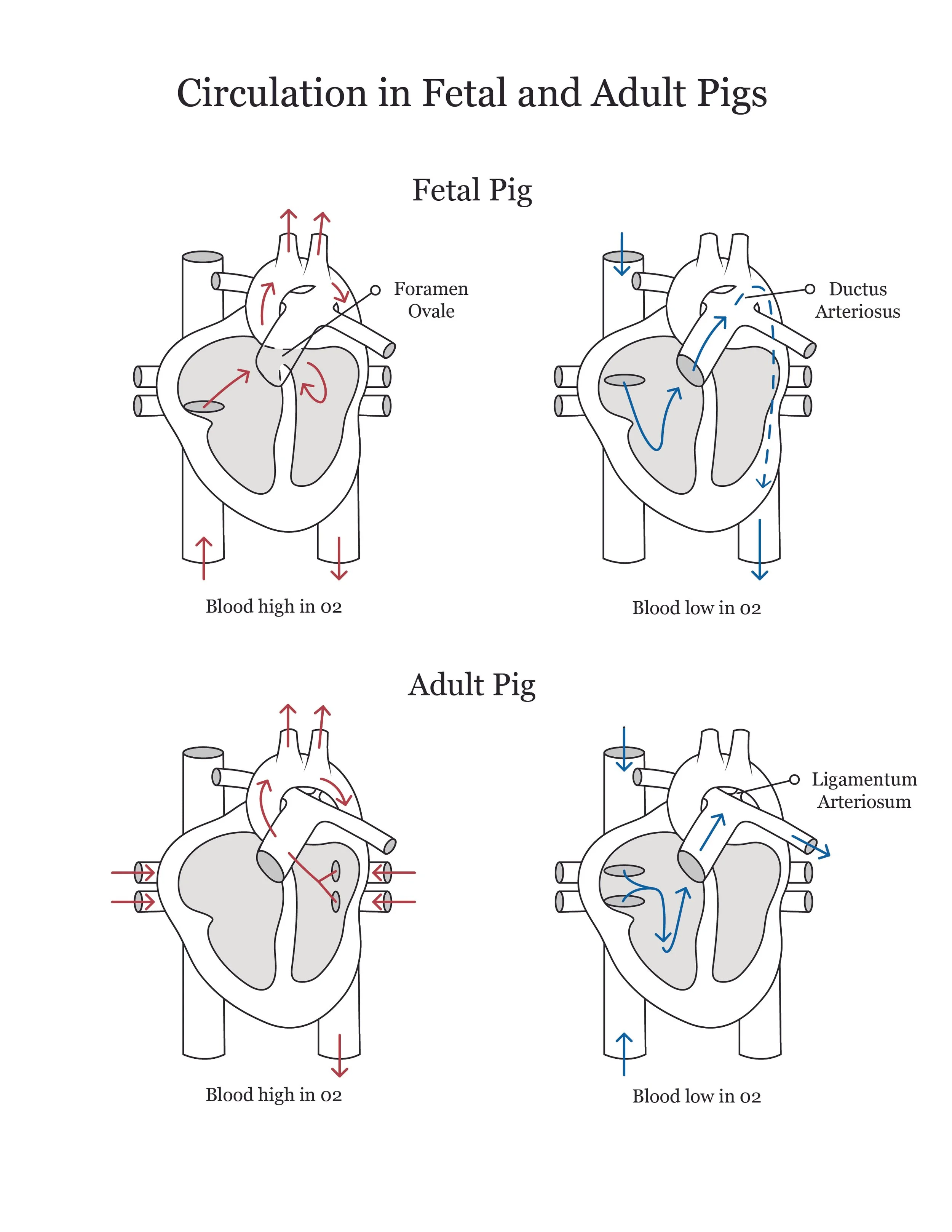
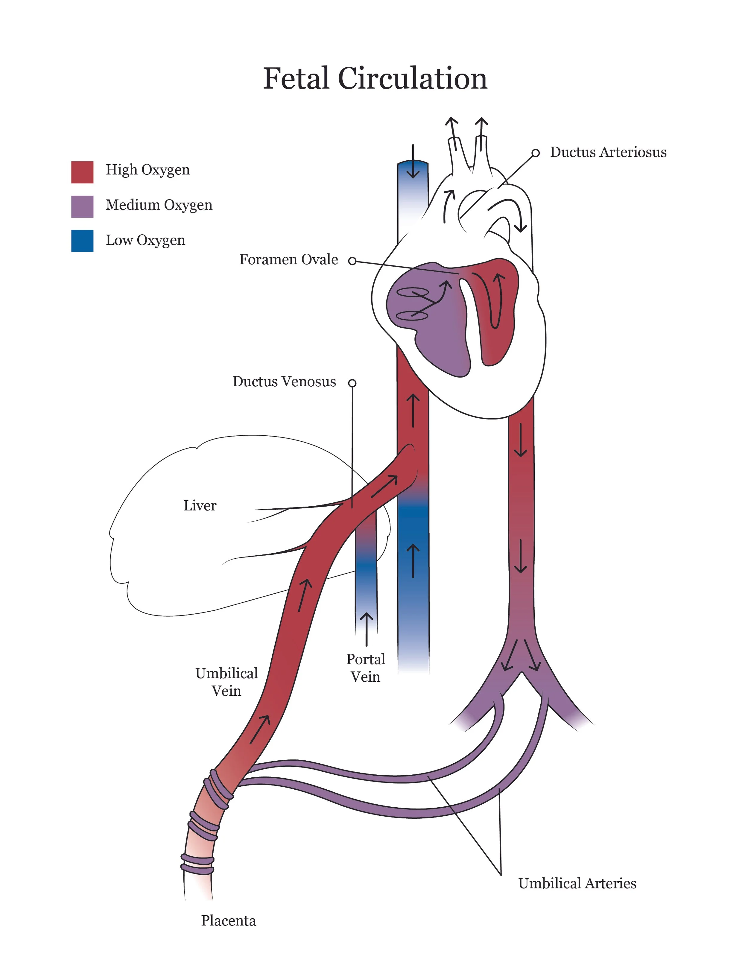
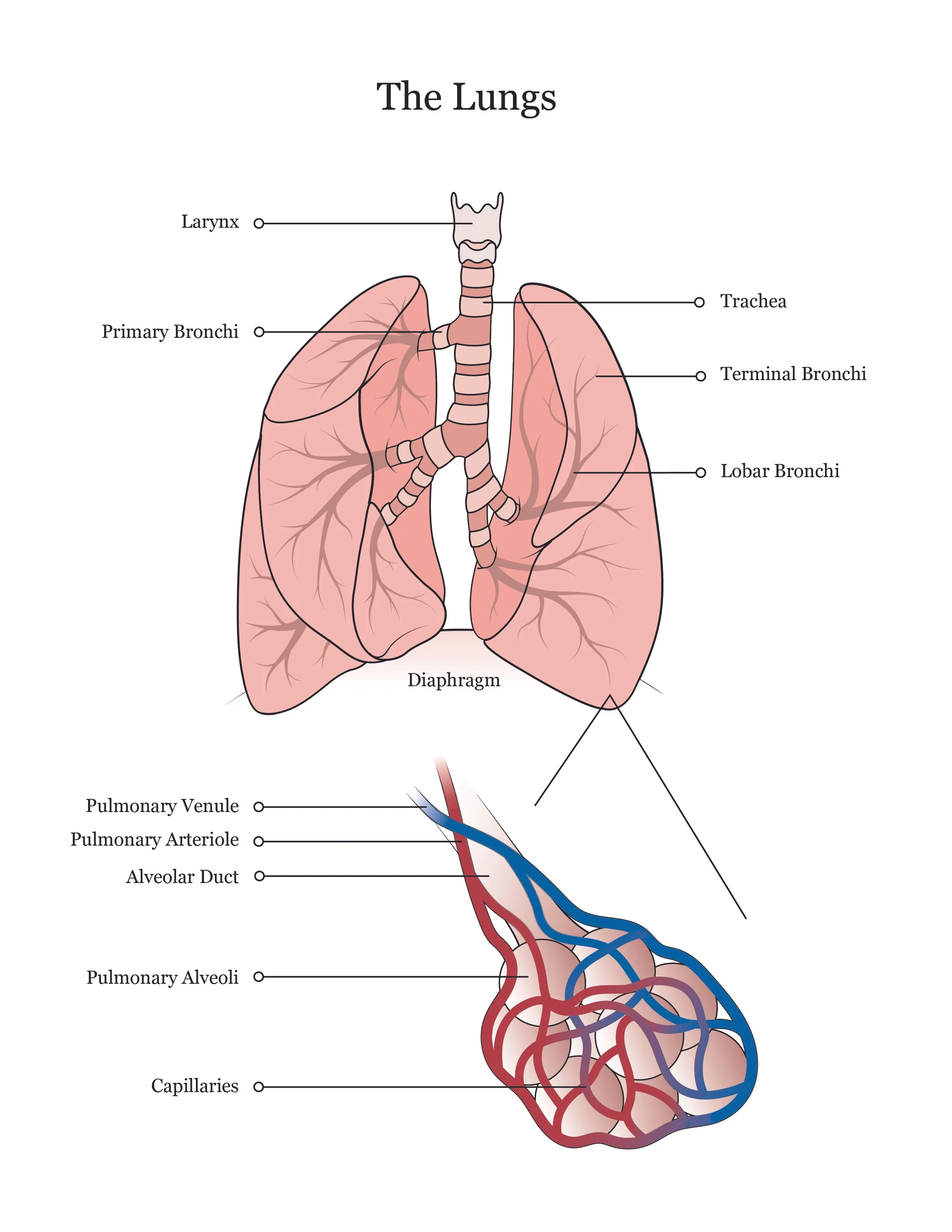
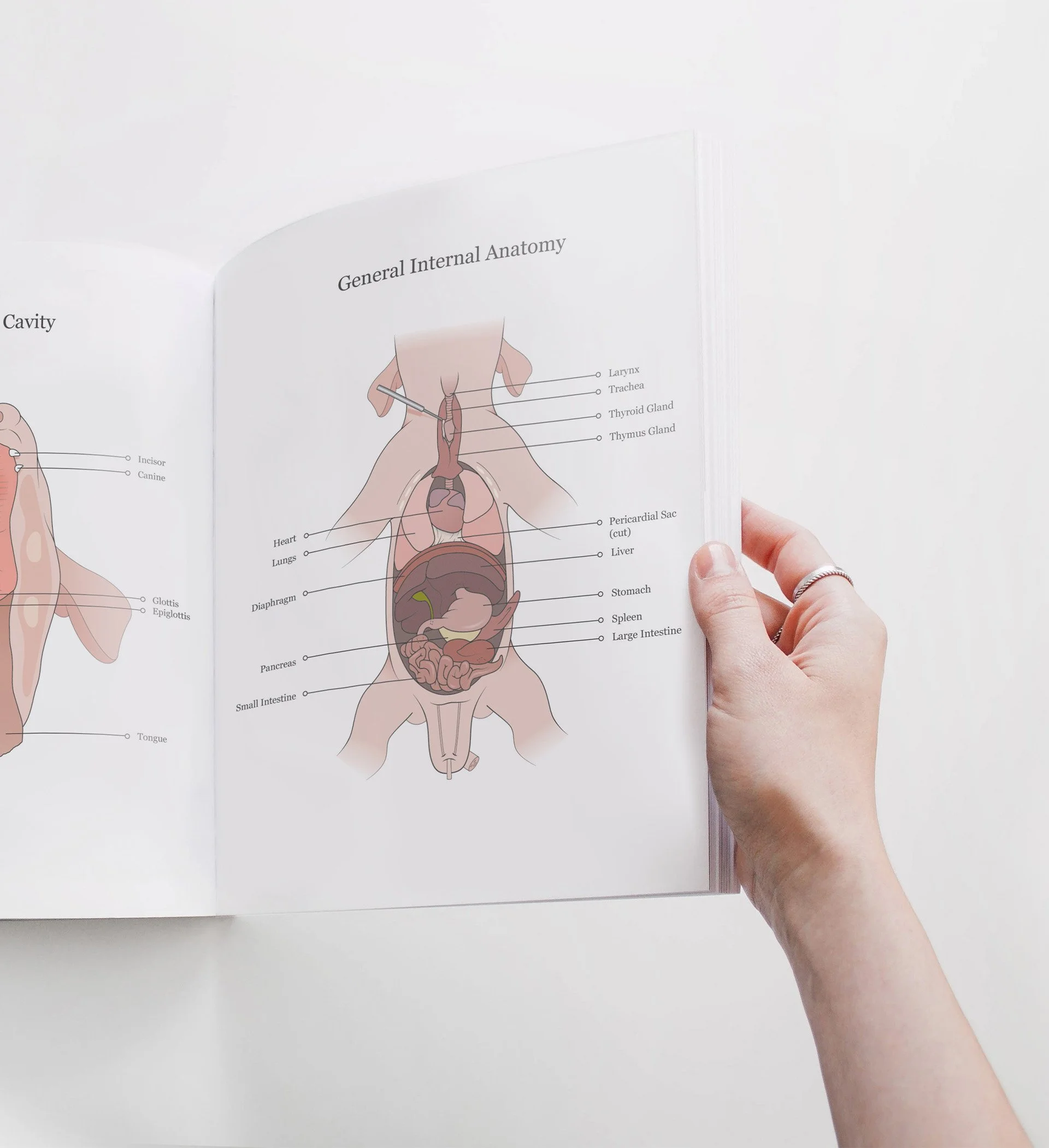
This project involved illustrating a series of diagrams for a fetal pig dissection manual, focusing on the digestive, urogenital, and arterial systems. I consolidated multiple reference sources and firsthand specimen observation into a cohesive visual system for instructional use. Illustrations were refined iteratively to ensure anatomical accuracy, consistency, and clarity for students.
Role Illustrator, Researcher
Project Type Educational Design
Context Academic
PROJECT BREAKDOWN
-
The manual had been assembled from mismatched sources over the years, so the visual language shifted from page to page. Repeated scanning had degraded details, leaving unclear shading in areas where precision mattered most. Instructors had flagged inaccuracies for years, but without a unified source file, students were left interpreting anatomy through diagrams that didn't always match what they saw on the table.
-
I sat through the course as a student, tracking where visual confusion actually happened, not just where the old diagrams looked outdated. Using published references and specimen observation, I rebuilt each illustration to standardize line work, shading, and labeling across. Every draft went through multiple iterations with the instructor for anatomical precision, then with students to confirm the diagrams matched what they were seeing.
-
Students gained one cohesive set of diagrams they could trust, replacing a patchwork that had caused confusion for years. The instructor now had a source file that could be corrected and expanded going forward, instead of relying on scans. More broadly, the project showed how illustration quality directly shapes comprehension in hands-on science education, where an unclear boundary can affect a student's understanding of anatomy itself.